Vein disease often develops gradually, with symptoms that can be easy to overlook until they become a serious concern. That’s why early detection is key. Identifying vein issues before they progress can prevent complications like chronic pain, swelling, and even life-threatening conditions such as deep vein thrombosis (DVT).
Diagnostic tools for vein problems begin with non-invasive techniques such as a physical exam and expand to more invasive measures like intravenous ultrasounds. The type of diagnostic tool one needs to monitor one's vein health depends on several factors, including the risk of vein disease and lifestyle factors.
In this blog post, you will learn about the most common diagnostic tools for vein disease and emerging technologies that will forever change how vein disease is detected and treated.
The Importance of Accurate Vein Problem Diagnosis
Accurate diagnosis of vein problems is essential for finding effective treatments and improving patient outcomes. To achieve these benefits, it is important to undergo early screening. Early detection can significantly reduce the risk of disease progression and complications.
However, using the correct diagnostic approach is crucial to maximize the advantages of early detection.
These approaches include:
- Physical examination (Including cough impulse and Brodie-Trendelenburg)
- Duplex ultrasound
- Doppler ultrasound
- Air plethysmography
- MR venography
- CT venography
- Near-infrared imaging and thermography
- Ankle-brachial index (ABI)
- Photoplethysmography
- Intravascular Ultrasound (IVUS)
Physical Examination Techniques for Vein Disease
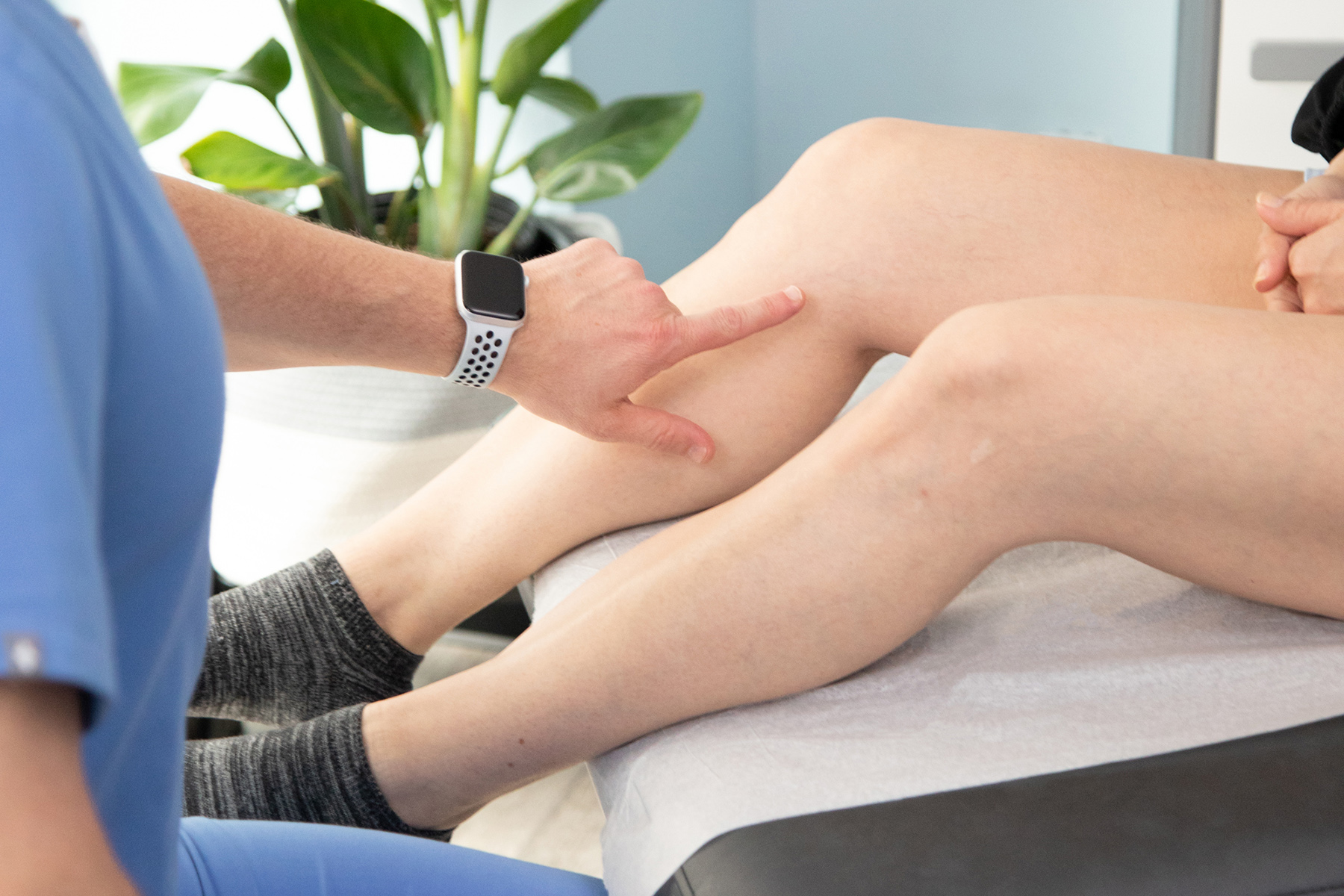
Before applying advanced diagnostics, a vein specialist will perform a visual examination of the area of concern to look for signs of swelling, warmth, skin changes, visible veins, and discoloration. They may also palpate the area to check for tenderness and pain.
In addition to a visual exam, your vein specialist may administer one of the following specialized tests:
- Cough Impulse: During this exam, the specialist will ask you to cough while they palpate your thigh to check for a palpable thrill, which could indicate venous reflux.
- Brodie-Trendelenburg: This test requires the specialist to empty your leg vein by elevating your leg and applying a tourniquet around your thigh. Then, you will stand up so the specialist can observe how quickly your veins fill with blood below the tourniquet.
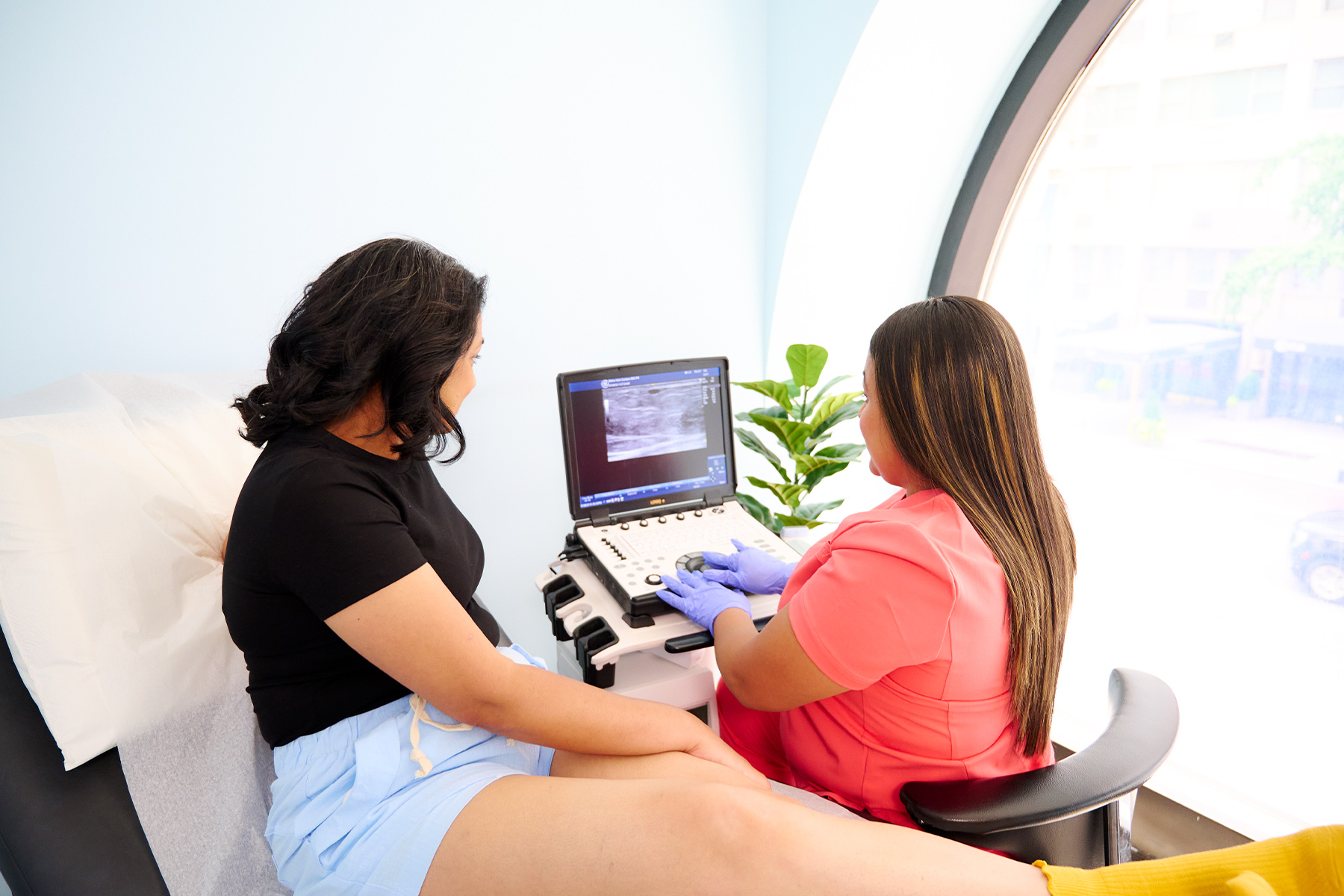
Duplex Ultrasound: The Gold Standard for Vein Disease Detection
A duplex ultrasound is the gold standard for detecting vein disease. This diagnostic procedure combines ultrasound imaging with Doppler sound to give the doctor an accurate view of the vein’s anatomy and blood flow. Your specialist will order a duplex ultrasound if they suspect a blood clot, varicose veins, or venous reflux.
Advanced Imaging Techniques
When duplex ultrasound is not enough to diagnose a vein issue, you might require advanced imaging such as an MR Venography, CT Venography, Near-Infrared Imaging, or Thermography.
- MR Venography: Utilizes magnetic resonance imaging (MRI) to view leg veins in detail.
- CT Venography: Uses computer tomography (CT) imaging with contrast to create a detailed image of the vein.
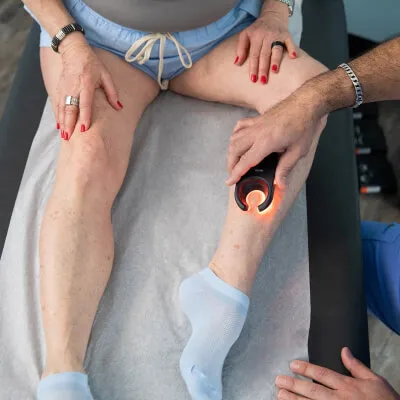
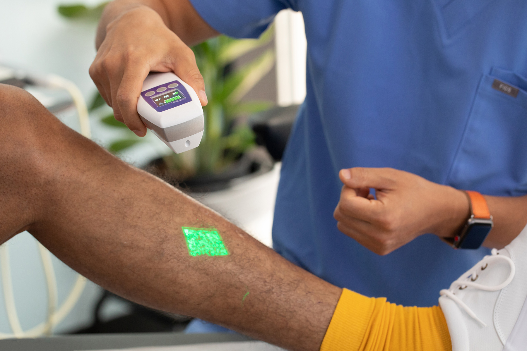
- Near-Infrared Imaging: Uses a handheld medical device that emits near-infrared light to view veins beneath the skin.
- Thermography: Uses infrared thermal imaging to assess variations in temperature between the veins, pinpointing poor blood flow and deep vein thrombosis.
Non-Invasive Vascular Tests for Vein Health
Noninvasive vascular tests provide information regarding blood flow and the overall health of your veins without using invasive techniques. Two commonly used tests for this purpose are the Ankle-Brachial index (ABI) and photoplethysmography.
- Ankle-Brachial Index (ABI): This test uses blood pressure measurements taken at the ankle and arm with a Doppler device and sphygmomanometer. The two results are calculated to reveal a systolic blood pressure ratio. The ratio is interpreted to reveal a normal or abnormal ABI indicating peripheral artery disease (PAD).
- Photoplethysmography: This test detects chronic venous insufficiency and venous reflux. It is performed by placing a small sensor on the skin and emitting infrared light. How much light is absorbed and reflected by the tissue underneath reveals the extent of CVI.
Photoplethysmography and Air Plethysmography Tests
If the doctor suspects you have a blood flow issue, they may order a photoplethysmography or air plethysmography.
- Photoplethysmography (PPG): A non-invasive procedure that uses infrared light to measure changes in blood volume within the veins.
- Air plethysmography (APG): Uses an air-filled cuff around the leg to measure venous function in varying movements dealing with elevation and dependency.
Both tests are useful in detecting chronic venous insufficiency, post-thrombotic syndrome, and varicose veins.
Vein Disease Classification Systems and Scoring
Tools like CEAP classification and the Venous Clinical Severity Score (VCSS) offer a standardized approach to diagnosing, treating, and monitoring the progression of diseases like venous reflux and chronic venous insufficiency (CVI).
- CEAP Classification: CEAP stands for clinical (signs and symptoms), etiology (cause), anatomy (of the vein), and pathophysiology (abnormalities). This framework is useful for assessing, diagnosing, and classifying vein disorders such as perforator incompetence.
- Venous Clinical Severity Score (VCSS): Like CEAP, VCSS uses a scoring system to assess, monitor and evaluate treatment progress. VCSS scores the impact of vein disease on the patient, monitors changes over time, measures the effectiveness of treatment, and compares the patient's progress with that of others in a similar group.
Specialized Diagnostic Procedures for Further Imaging
Specialized diagnostic procedures, such as venography and intravascular ultrasound, are reserved for cases where noninvasive tests are inconclusive or further imaging is needed to reveal the extent of a vein disease.
- Venography: A specialized test that combines X-ray technology and contrast to assess vein obstructions and abnormalities in cases where other tests are inconclusive.
- Intravascular Ultrasound (IVUS): Checks for blockages and guides vein surgeons during complex treatments by way of a tiny insertable ultrasound probe.
Emerging Technologies in Vein Diagnostics
Emerging health monitoring technology allows patients to monitor their vein health outside a specialist's office. Using artificial intelligence, they can analyze patterns in vein diagnostics like ultrasounds or venography to detect abnormalities. Similarly, wearable devices such as smart compression garments or biosensors provide vein health data, allowing patients to monitor venous blood flow, pressure, and swelling.
Metro Vein Centers offers the latest state-of-the-art tools and vein treatment technology. Schedule a free diagnostic vein evaluation with us today.
Frequently Asked Questions
How accurate is duplex ultrasound in diagnosing deep vein thrombosis?
According to the National Blood Clot Alliance, duplex ultrasound successfully identifies 60 to 70% of all calf vein thrombosis.
Can vein problems be diagnosed without invasive procedures?
Yes. Several noninvasive diagnostic techniques, such as CT scans and ultrasound, can accurately detect leg vein issues.
What's the difference between CEAP classification and Venous Clinical Severity Score?
CEAP categorizes various aspects of chronic venous disease (CVD), accurately describing its characteristics. VCSS scores the severity of those characteristics.
How often should someone with a family history of vein problems get diagnostic tests?
If you have a family history of vein disease, you should have a vein screening annually or at least every two years. Your vein specialist will recommend a screening schedule that is best for you.
Are there any new diagnostic tools being developed for vein disease detection?
Yes, tools for vein disease detection continue to advance through innovation, including the use of artificial intelligence. Several new studies highlight the use of portable vein analyzers and artificial intelligence-powered ultrasounds to diagnose vein abnormalities and blood clots.

Dr. Philip LoPresti
Meet Dr. Philip LoPresti, DABVLM, FACS, a board-certified vein specialist and surgeon with over 20 years of experience. Schedule an appointment with him in Queens, NY today.
Meet Dr. Philip LoPrestiTrusted insight from the nationally accredited, board-certified vein doctors at Metro Vein Centers.